Practice closedClosed
Practice closedClosed
Saving the menisci: suture rather than resection
"The meniscus is an irreplaceable shock absorber. My philosophy, in line with SFA recommendations, is to preserve it systematically whenever possible, repair rather than resection. This protects your knee against osteoarthritis."
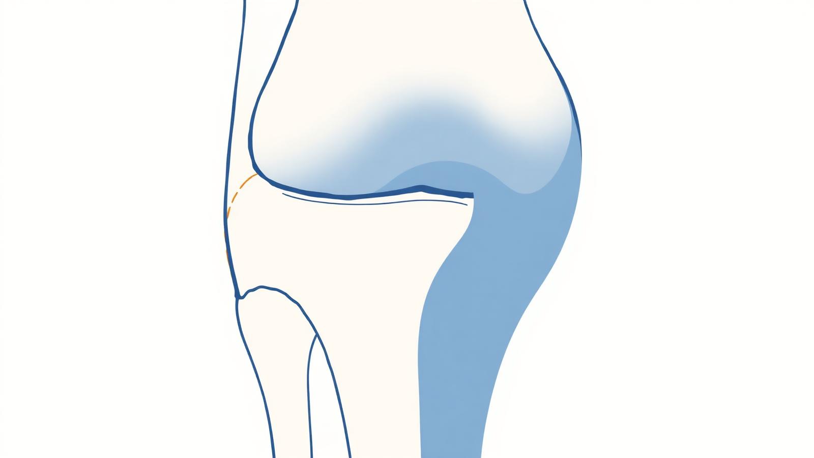
Meniscal tears are common, occurring at a rate of about 9 per 10,000 in men and 4.2 per 10,000 in women. For a long time, the surgical response was to remove the damaged meniscus. We now know this was a mistake: the meniscus is an irreplaceable shock absorber, and its removal accelerates cartilage wear and osteoarthritis. The goal of every modern practitioner, and Dr Charousset's strong conviction, is now to save the menisci whenever possible. Understanding the anatomy, biomechanics and evolution of the menisci is essential to analyze tears and choose their treatment.
The most important distinction today is between stable and unstable tears, since their treatment differs radically. A stable meniscal tear is often degenerative, related to joint aging, age, but also to occupational or sporting activity. It is frequently accompanied by cartilage lesions of the femorotibial compartment. It can cause knee pain, sometimes with intra-articular fluid effusion. An unstable meniscal tear is most often traumatic: it detaches a fragment of meniscus of varying size, causing locking of the knee accompanied by pain.
Meniscal tears vary greatly in shape and location. They may be fissures, more or less mobile flaps, or large displaced fragments called bucket-handle tears, often responsible for acute knee locking (the knee remains stuck in flexion, impossible to straighten). Tears can be vertical, horizontal or radial. The medial meniscus is mainly affected in its posterior part, with progressive extension towards the middle part. The lateral meniscus is mainly affected in its middle part, with possible extension forward or backward.
MRI is the key test to diagnose a lesion, tear or rupture of the meniscus. A protocol including T2-weighted sequences is particularly important to properly analyze the meniscus. MRI defines the shape of the lesion, its location and its stable or unstable nature, information that directly determines the therapeutic choice. CT arthrography or MR arthrography can complete the workup in complex cases or for a previously operated meniscus.
Stable or degenerative tears are primarily managed with medical treatment, without surgery. This combines joint rest, analgesics and cryotherapy (cold application). In certain cases, when associated cartilage lesions are present, Dr Charousset offers PRP injections: this regenerative medicine technique, of which he is a pioneer in France, acts on pain and on the cartilage environment. Many degenerative meniscal tears improve this way without surgery.
Surgery is primarily aimed at unstable meniscal tears responsible for knee locking. Dr Charousset's philosophy, in line with the recommendations of the French Society of Arthroscopy (the 'Save the Menisci' campaign), is to preserve the meniscus as much as possible. Two options are available, performed under arthroscopy: meniscus repair (suture), which reinserts and repairs the torn meniscus to keep it, or minimal trimming of an unstable flap, which removes only what is strictly necessary while preserving as much meniscal tissue as possible. Wide resection, once systematic, is now avoided as much as possible.
The possibility of repairing a meniscus depends on its blood supply, since only vascularized tissue can heal. Three zones are described, from the periphery towards the center of the knee: the red-red zone (peripheral, well vascularized, close to the joint capsule), the red-white zone (intermediate) and the white-white zone (central, non-vascularized). Meniscus repair is only possible in vascularized zones, meaning close to the capsule (red-red zone and, to a lesser extent, red-white). A lesion in the white-white zone, non-vascularized, will not heal and requires economical trimming.
Arthroscopic meniscal surgery is performed as outpatient surgery. Recovery depends on the procedure performed: after simple trimming, recovery is fast (a few weeks); after meniscus repair, protection is longer (the repaired meniscus must heal), with a gradual return to activity over several months and adapted rehabilitation. The choice to preserve the meniscus, even if it requires a slightly longer recovery, durably protects the knee against osteoarthritis. It is an investment in the future of the joint.
Some videos contain surgical footage that may be disturbing for sensitive viewers.
Qu'est-ce qu'une lésion méniscale ?
Rappels d'anatomie et mécanismes lésionnels.
Le catalogue des lésions méniscales (anses de seau)
Panorama des différentes formes de rupture.
Suture méniscale sous arthroscopie et injection de PRP
Préservation méniscale associée à un renfort biologique.
Le Dr Charousset vous reçoit à l'IOAPC, 23 rue de Courcelles, Paris 8e.