Practice closedClosed
Practice closedClosed
The tests used to examine bone, menisci, ligaments and cartilage
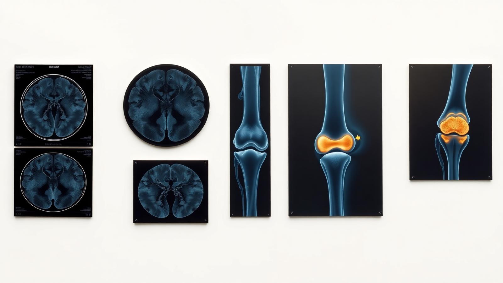
"Choosing the right imaging test avoids unnecessary prescriptions. Targeted, high-quality imaging allows a precise diagnosis to be made from the first consultation."
The knee contains structures of very different natures, bone, cartilage, menisci, ligaments, tendons, that no single test can explore perfectly. Knee imaging therefore relies on several complementary techniques, each suited to a particular type of structure. Dr Charousset chooses the most relevant test based on the suspected condition, in order to obtain a precise diagnosis without multiplying unnecessary tests.
X-ray is the basic test for examining bony structures. It reveals fractures, osteoarthritis (through joint space narrowing, osteophytes), limb axis abnormalities and calcifications. Performed weight-bearing (standing), it assesses the impact of osteoarthritis on knee alignment. Simple and quick, it is often the first test ordered.
Ultrasound is useful for examining tendon disorders and peri-articular soft tissue. Non-invasive and dynamic, it visualizes tendons (patellar, quadriceps), detects effusions, cysts and inflammation. It also guides injections and PRP procedures.
CT arthrography involves performing a CT scan after injecting a contrast agent into the joint. This agent 'molds' all internal structures, allowing detailed study of the cartilage, the deep surface of the ligaments, the joint capsules and the menisci. It is particularly precise for cartilage analysis and for visualizing the menisci, which are invisible on a plain X-ray.
MRI (Magnetic Resonance Imaging) is the reference test for examining the soft tissues of the knee. Without injection or radiation, it precisely explores the cartilage, the menisci, the collateral ligaments (medial and lateral), the cruciate ligaments (anterior and posterior), as well as the patellar and quadriceps tendons. It is the key test for diagnosing a meniscal tear or a ligament rupture. A protocol with T2-weighted sequences is particularly important for meniscal analysis.
MR arthrography combines MRI with the injection of an intra-articular contrast agent. It combines the fine soft-tissue analysis of MRI with the precision brought by the contrast agent, particularly for complex meniscal tears, subtle cartilage lesions, or the analysis of a previously operated meniscus.
An essential point to understand: the menisci are not calcified like bone, so they are invisible on a plain X-ray. Two tests allow them to be visualized. CT arthrography, using the injected contrast agent, which is also very useful for studying cartilage. And MRI, without injection, which is also valuable for studying the knee ligaments, particularly the anterior cruciate ligament. The choice between these two tests depends on the type of lesion being sought: cartilage lesion, ligament lesion, or isolated meniscal lesion.
Dr Charousset or your doctor will order one or another of these tests depending on the suspected diagnosis and any associated lesion to be investigated:
Well-chosen, high-quality imaging allows a precise diagnosis from the consultation.
Le Dr Charousset vous reçoit à l'IOAPC, 23 rue de Courcelles, Paris 8e.