Practice closedClosed
Practice closedClosed
Cartilage wear beneath the kneecap
"Patellar chondropathy is treated first with rehabilitation. When pain persists, PRP is an effective option before considering realignment surgery."
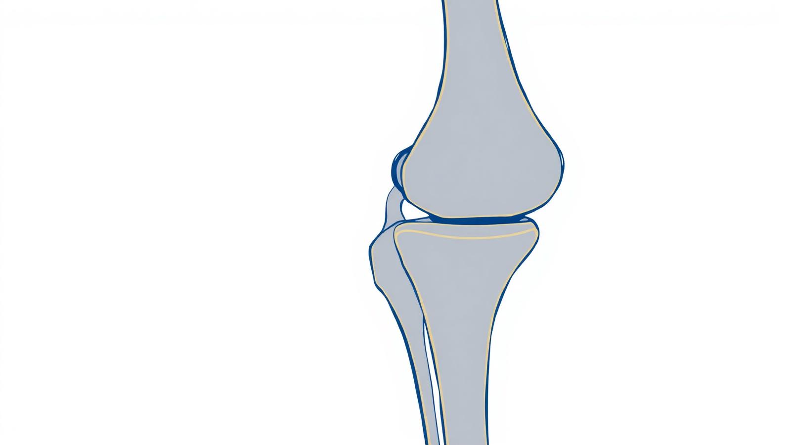
Patellar chondropathy refers to damage to the cartilage that covers the back surface of the kneecap and the groove of the femur in which it glides (the trochlea). This cartilage, which should allow perfectly smooth gliding of the kneecap during flexion-extension movements, softens, cracks and then wears away. Chondropathy is a common cause of knee pain, particularly in young, athletic people and in women.
The kneecap and the femoral trochlea form the patellofemoral compartment, one of the three compartments of the knee. With every knee flexion, the kneecap is pressed against the femur with a force that increases considerably with the flexion angle: climbing stairs, squatting or standing up from a low seat subject this cartilage to high pressures. This explains the very specific nature of chondropathy pain.
Chondropathy can be primary (cartilage wear with no obvious anatomical cause) or favored by a misalignment of the extensor mechanism: a kneecap that does not glide perfectly in its groove (poor patellar tracking), a trochlea that is too flat, a kneecap positioned too high, or poor muscular balance of the quadriceps. Repeated microtrauma from sport, sequelae of a kneecap dislocation, or being overweight are also contributing factors.
Patellar chondropathy pain is characteristic: it is located at the front of the knee, around or behind the kneecap. It is triggered by situations that load the patellofemoral compartment: climbing and especially descending stairs, squatting, prolonged sitting with the knee bent (the well-known 'movie theater sign': pain after sitting for a long time). Cracking (crepitus) may be felt, and sometimes a catching sensation during movement.
Clinical examination reveals pain on palpation and mobilization of the kneecap, and reproduces the pain during maneuvers that load the cartilage. X-ray, with specific views (axial view of the kneecap), analyzes patellar tracking and trochlear morphology. MRI or CT arthrography precisely assess the state of the cartilage (depth and extent of wear), classified into several grades of increasing severity.
Treatment is primarily medical and prolonged. Its cornerstone is rehabilitation: targeted quadriceps strengthening (particularly its medial part) to improve patellar tracking, combined with stretching and proprioceptive training. Orthotic insoles are added in cases of static disorders, along with activity modification and weight loss if needed. In persistently painful forms, Dr Charousset offers PRP injections into the knee: this regenerative technique has a favorable effect on cartilage and pain, and represents a valuable option before considering surgery.
Surgery is only considered in case of failure of well-conducted medical treatment and when there is a correctable anatomical disorder. Under arthroscopy, Dr Charousset can perform cleaning and trimming of unstable cartilage lesions (debridement). When chondropathy is linked to confirmed poor patellar tracking, a realignment procedure of the extensor mechanism (such as an osteotomy of the tibial tuberosity, which repositions the attachment point of the patellar tendon) may be indicated to correct the mechanical cause.
Recovery depends on the procedure performed. After a simple arthroscopic procedure, recovery is relatively quick with rehabilitation focused on strengthening and patellar tracking. After a bony realignment, protection is longer while healing occurs. In all cases, rehabilitation remains the determining factor for the result, and the patient plays a major active role in recovery through regular muscle work.
Le Dr Charousset vous reçoit à l'IOAPC, 23 rue de Courcelles, Paris 8e.