Practice closedClosed
Practice closedClosed
Understanding the most complex joint in the body
"A knee is a precision mechanism. My role is to preserve its natural structures as much as possible, menisci, ligaments, cartilage, and to precisely restore its anatomy when surgery is necessary."
The knee is the largest joint in the human body, and one of the most heavily used. With every step it bears several times body weight; with every run, jump or change of direction it absorbs considerable stress. Its particularity is having to reconcile two contradictory requirements: great stability to support these loads, and good mobility to allow flexion, extension and slight rotation. This balance relies on the harmony of several structures working together.
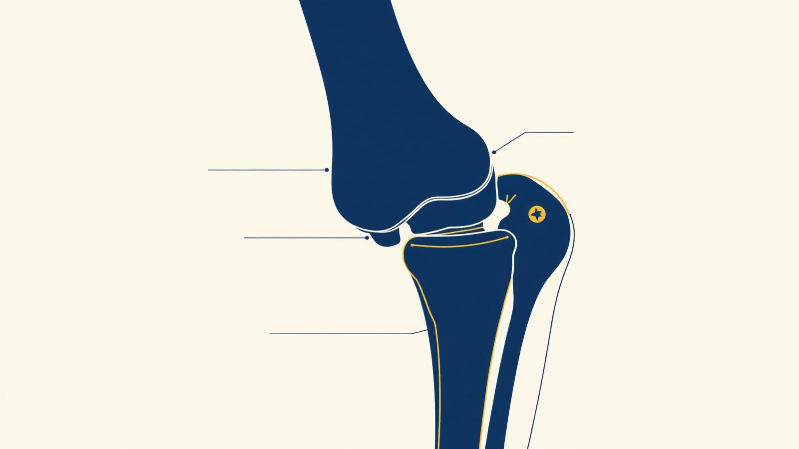
The knee brings together three bones. The femur, the thigh bone, whose lower end forms two rounded prominences called condyles. The tibia, the main bone of the leg, whose upper end, the tibial plateau, receives the femoral condyles. And the patella (kneecap), a small bone at the front, embedded in the quadriceps tendon, which slides in a groove of the femur (the trochlea) during flexion-extension movements. These three bones define three compartments: medial, lateral and patellofemoral.
The contact surfaces between these bones are covered with articular cartilage, a smooth and resistant tissue that allows frictionless gliding and absorbs shocks. This cartilage, which has no blood vessels or nerves, does not heal spontaneously when injured, which explains the potential severity of its wear. Its progressive breakdown is the origin of osteoarthritis.
Between the femur and the tibia sit two menisci, medial and lateral, true shock absorbers of the joint. They distribute loads, improve the congruence between bone surfaces and contribute to stability.
Knee stability is ensured by four main ligaments, cord-like structures that connect the femur to the tibia and guide movement while preventing abnormal displacement:
The meniscus is the shock absorber of the knee. There are two: one medial and one lateral. Crescent-shaped, they adapt to knee movements. The medial meniscus is shaped like an open C; it is attached to the medial collateral ligament along its entire length, which makes it more stable but also more vulnerable to shear forces created by knee rotation. The lateral meniscus, more closed (almost O-shaped), is not attached to the capsule along its entire length and remains separate from the lateral collateral ligament: it is therefore more mobile but less stable. The two menisci are connected to each other and to the cruciate ligaments, with which they work in harmony, following the movements of the femur and tibia during flexion and extension.
Understanding this anatomy helps understand its pathologies. A torn cruciate ligament, a split meniscus, a worn cartilage: each of these injuries unbalances the fine mechanics of the knee and affects the other structures. This is why Dr Charousset places great importance on preserving these elements as much as possible, particularly the menisci, and on precisely restoring the anatomy when surgery is necessary.
Le Dr Charousset vous reçoit à l'IOAPC, 23 rue de Courcelles, Paris 8e.