Practice closedClosed
Practice closedClosed
When the kneecap slips out: dislocation and subluxation
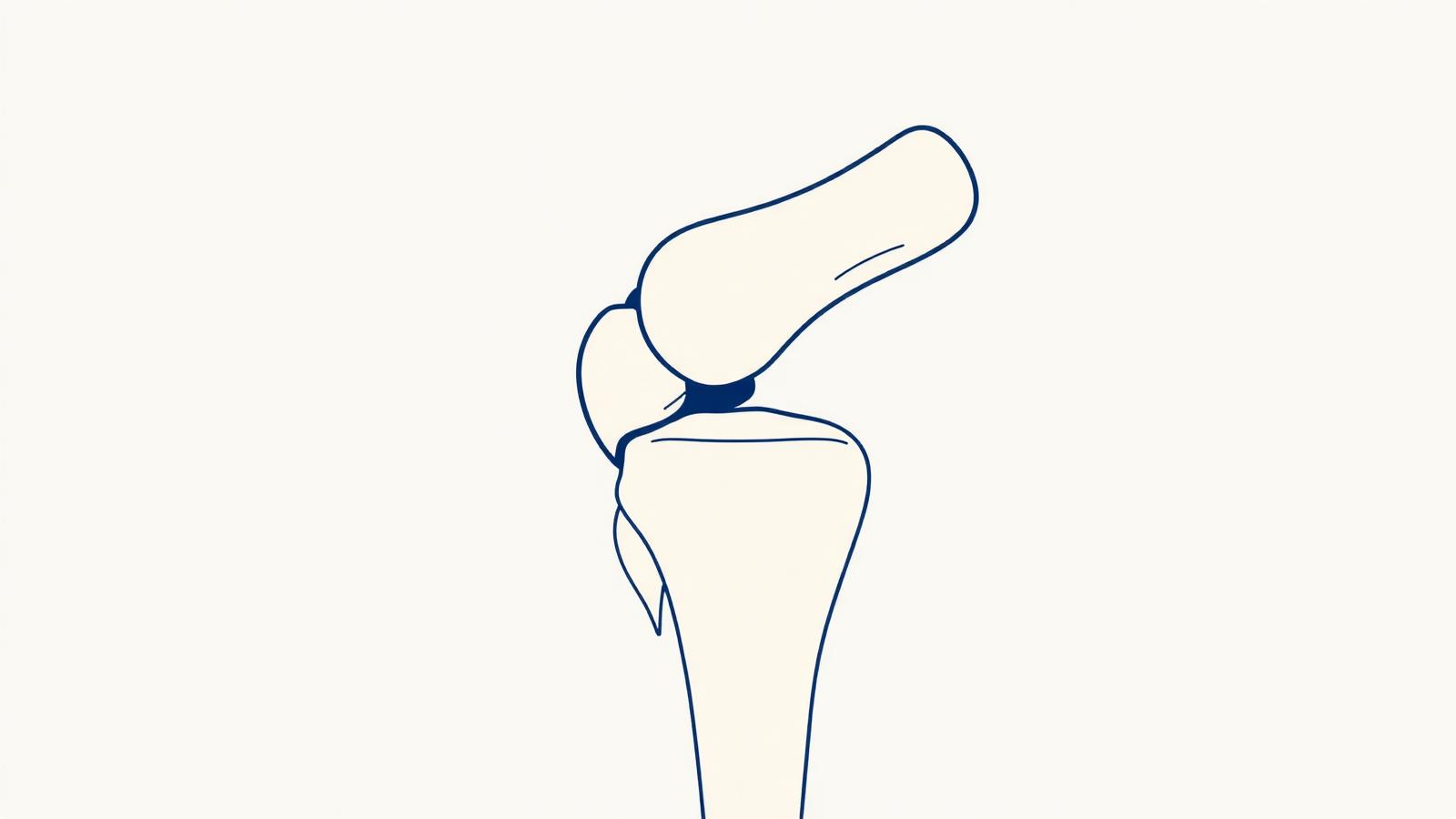
"Patellar instability is treated on a case-by-case basis. A complete morphological workup, X-rays, CT, MRI, precisely identifies the anatomical factors, and surgery only corrects what needs correcting."
Patellar instability refers to the tendency of the kneecap to come out of the femoral groove (the trochlea) in which it should normally glide. Unlike chondropathy, which concerns cartilage wear, instability concerns abnormal positioning of the kneecap, which shifts abnormally outward. This condition preferentially affects adolescents and young adults, more often women, and frequently relies on predisposing anatomical features.
Patellar dislocation is a complete displacement: the kneecap comes out of the trochlea, almost always outward, during a flexion-rotation movement or trauma. It is a sudden and painful episode, sometimes with spontaneous reduction. Subluxation is an incomplete and transient displacement: the kneecap shifts partially then returns, leaving a feeling of instability, catching or apprehension. As with the shoulder, a first dislocation carries a risk of recurrence, which is higher when predisposing anatomical factors are present.
Several anatomical features predispose to patellar instability and must be looked for, as they guide treatment:
The patient reports episodes of the kneecap slipping out, or a feeling of instability and apprehension, particularly during pivoting or flexion movements. After a true dislocation, swelling and pain are observed on the inner side of the knee (where the MPFL tore). Between episodes, discomfort, cracking and apprehension with certain movements may persist.
Diagnosis is clinical, but the essential part of management is the complete morphological workup, indispensable before any decision. It combines specific X-rays, a CT scan (measuring TT-TG distance, analyzing the trochlea) and an MRI (state of the MPFL, cartilage). This workup precisely identifies the anatomical factors involved in each patient, allowing a tailored treatment adapted to the abnormalities actually present.
After a first dislocation without severity factors, treatment can be medical: brief immobilization, then intensive rehabilitation focused on muscle strengthening (quadriceps, particularly its medial part) and proprioceptive training to stabilize the kneecap. This management allows many patients to avoid recurrence.
In case of recurrence or marked anatomical factors, surgery is offered, adapted to the abnormalities identified during the workup. MPFL reconstruction restores the torn internal restraint, replacing the medial patellofemoral ligament with a graft. Osteotomy of the tibial tuberosity (TTA) repositions the attachment point of the patellar tendon to correct an excessive TT-TG distance and realign the extensor mechanism. These techniques can be performed alone or combined depending on each patient's workup. The goal is to durably recenter the kneecap.
Recovery depends on the procedures performed. After MPFL reconstruction, protection is needed while the graft integrates; after an osteotomy, while bone healing occurs. In all cases, progressive rehabilitation restores range of motion, strength and stability. Return to sport is supervised and adapted to each case. Correction of anatomical factors gives good long-term results on patellar stability.
Le Dr Charousset vous reçoit à l'IOAPC, 23 rue de Courcelles, Paris 8e.